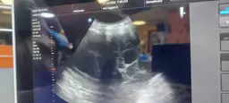
Empyema
Empyema:
- Empyema refers to the presence of infected pus within the pleural space, typically as a complication of pneumonia.
- In lung ultrasonography, empyema appears as a complex fluid collection with internal septations and debris. The fluid may also have heterogeneous echogenicity, and there may be loculations within the pleural space.
- Ultrasonography can also show pleural thickening and irregular pleural surfaces in cases of empyema.
- Ultrasonography can assess the size and extent of the empyema, guide thoracentesis or chest tube placement, and monitor the response to treatment.
Bronchopleural Fistula:
- A bronchopleural fistula is an abnormal communication between the bronchial tree and the pleural space, often resulting from conditions such as lung abscess, necrotizing pneumonia, or post-surgical complications.
- In lung ultrasonography, a bronchopleural fistula may appear as an air-fluid level within the pleural space, with dynamic air movement visualized during respiration.
- Ultrasonography can also show the presence of air bubbles or air-fluid interfaces within the pleural space, which are characteristic findings of a bronchopleural fistula.
- Ultrasonography could be used to identify the location and size of the bronchopleural fistula, monitor changes in its characteristics over time, and guide interventions such as chest tube placement or surgical repair.